July/August Pearl #3: Fleck Sign
Pearl: Fleck Sign - small boney fragment seen in the space of the 1st and 2nd metatarsal that is associated with a Lisfranc ligament avulsion
Pearls by Drs. Grass (PGY-3), Roman (PGY-1), and Thompson (PGY-1).
Originally described by Myerson et al. in a series of tarsometatarsal joint fracture dislocations, this finding is felt to be present in 90% of patients with these injuries, more commonly referred to as LisFranc Injuries.
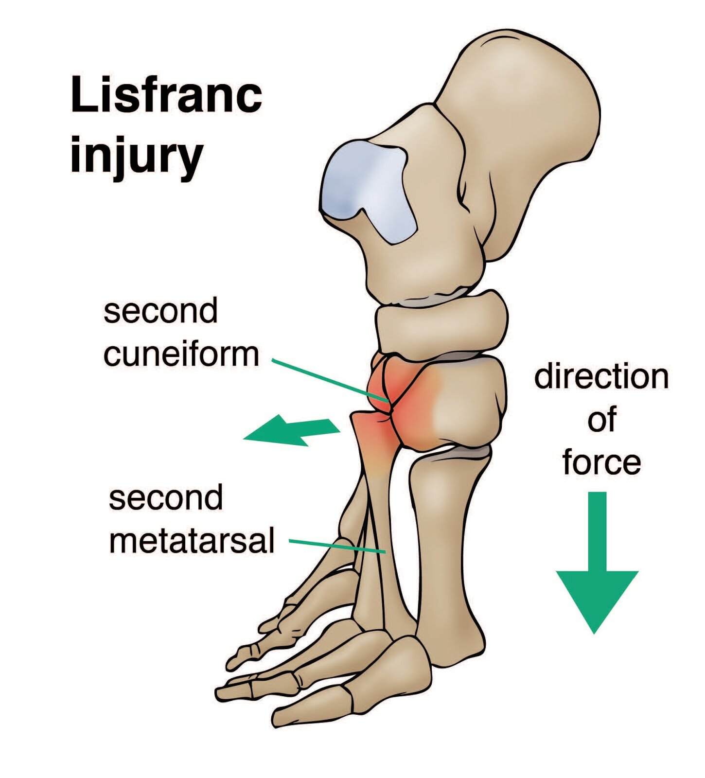
Lisfranc injuries are a spectrum of injuries from a simple sprain to complete disruption of the tarsi-metatarsal joints in the mid foot, that typically occur at the base of the 2nd metatarsal. The Lisfranc ligament itself is a strong band that attaches from the medial cuneiform to the 2nd metatarsal base and is crucial to stability of the midfoot. This injury most typically occurs from high force injury, however it can occur in low velocity injuries. In fact, it is the low velocity injuries with suspicious exam findings that should make one suspicious for this type of injury.

Mechanism
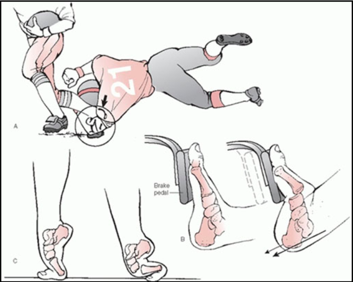
Usually a high force mechanism with plantar flexion + axial loading or external rotation.
common injuries (see picture below): car accident while slamming on the break, football/soccer being tackled from behind with plantar flexion, falling of a horse with foot stuck in the stirrup…
Diagnostics:
Imaging: typically done via X-ray
Adding on oblique view helps eliminate metatarsal overlap

Normal: note the alignment of the metatarsals and the cuneiform.

Abnormal
Left pane: circle shows widening >2mm between 1st and 2nd metatarsal concerning for unstable Lisfranc ligament injury (circle).
Middle pane: fracture of second metatarsal (arrow) and cuboid fracture (circle).
Right pane: no fractures, but rather a complete dislocation of the entire midfoot (box).

Subtle injury: weight bearing views (with adequate analgesia) can be obtained for high suspicion injuries with negative initial injuries. The picture on the right (arrowhead) shows widening of the 1st and 2nd metatarsal spaces with weight bearing images. Compare to the left without weight bearing showing what was read as normal (arrow).
Of note: CT and MRI might be indicated with negative injuries.
- Tos et al. is a small retrospective analysis showing 12% of negative or equivocal weight bearing X-ray films had a positive CT scan.
Disposition
Most will require orthopedic consultation and surgery. Those that don’t have widening of the 1st and 2nd metatarsal or los of arch height on lateral x ray with a simple sprain of the ligament can be discharged with a posterior slab splint and orthopedics follow up.

Recent Case:
Mechanism: high speed MVC of restrained driver. Note the metatarsal fractures, widening of the 1st and 2nd metatarsal, and fleck sign (arrows). Disposition: orthopedics took this patient to the OR (post op on right).
Blog Post: John Schneider MD PGY-4
#FOAM:
Resources:
Kim, D. H., Berkowitz, M. J., & Hrutkay, J. M. (2017). "Fleck Sign": Traumatic Avulsion Fracture of the Medial Cuneiform by Anterior Tibialis Tendon. International Journal of Foot and Ankle, 1(1). doi:10.23937/ijfa-2017/1710001
Myerson MS, Fisher RT, Burgess AR, Kenzora JE (1986) Fracture dislocations of the tarsometatarsal joints: End results correlated with pathology and treatment. Foot Ankle 6: 225-242.
Tos P, Titolo P, Chirila NL, Catalano F, Artiaco S. Surgical treatment of acute fingernail injuries. J Orthop Traumatol. 2012;13(2):57–62. doi:10.1007/s10195-011-0161-z