A 39 y.o. woman persents with peritonitis, she has a history of a peritoneal inclusion cyst which has been drained by IR.
Her cyst is shown from 3/19 and her CT from 6.5 prior to the IR drain placement

cyst seen on US 3/19

CT prior to cyst drainage.
The cyst was drained on 6/5 and alcohol was instilled. They were unable to replace a drained after several tries because the cyst had collapsed. When she returned with peritonitis the cyst was no longer visible on CT , there was no free air and no perforation noted with oral contrast on CT. She was taken to the OR by general surgery . What did they find?
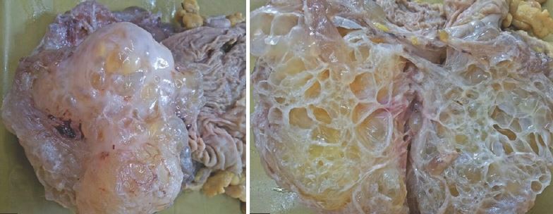
Our patient was found to have a 10 cm tubovarian abscess on the L. It was not diagnosed on CT scan possibly because of abnormalities from the collapsed inclusion cyst and possibly because CT is poor for defining pelvic structures. It is not clear if the cyst drainage caused inadvertent puncture of bowel or the cyst causing peritonitis.
peritoneal inclusion cysts caused by adhesions to the ovary.
A peritoneal inclusion cyst is usually caused by accumulation of ovarian fluid that is trapped by adhesions to the ovary caused by previous inflammation. Previous inflammation thickens the peritoneum and makes it more difficult to absorb fluid. Because the source of the fluid is ovarian, cysts are rare in males but have been reported in Crohn’s disease where there is chronic inflammation.
Conservative treatment of a peritoneal inclusion cyst is recommended because after surgical resection 30-50% recur. Oral contraceptives can be used to suppress ovulation, thus decreasing the formation of ovarian fluid trapped by adhesion and aspiration of the cysts can often be done transvaginally..
Our patient underwent a a L salpingo-oophorectomy and received doxy and flagyl . Her pain improved and she was discharged.
Jain, K. Imaging of peritoneal inclusion cysts. American Journal of Roentgenology. 2000 June Vol 174(6).
Ross M, Welch W, Scully R. Multilocular peritoneal inclusion cysts . Cancer 1989;64:1336-1364.
Sohaey R, Gardner T, Woodward P, Peterson C. Sonographic diagnosis of peritoneal inclusion cyst. J Ultrasound Med 1995;14:913-17.
Spriggs D, Melamed A, Weier L, Safdar N. A 24 y.o. woman with a pelvic mass. Case 18-2019. NEJM 2019
380:2361-9.
THE DIFFERENTIAL OF A PELVIC MASS INCLUDES:
Pregnancy and physiologic masses-endometriomas, cysts
Infections-diverticulitis, appendicitis, tuberculosis
Benign tumors-cystadenomas of the ovary or ovarian epithelial tumors
Cancer-familial tumors (BRCA or Lynch syndrome), high grade serous tumors and endometroid tumors(associated with CA-124), germ cell tumors.