A 23 y.o, presents to the ED with carpopedal spasms, you notice something unusual on exam of his skin.
What condition does he have?
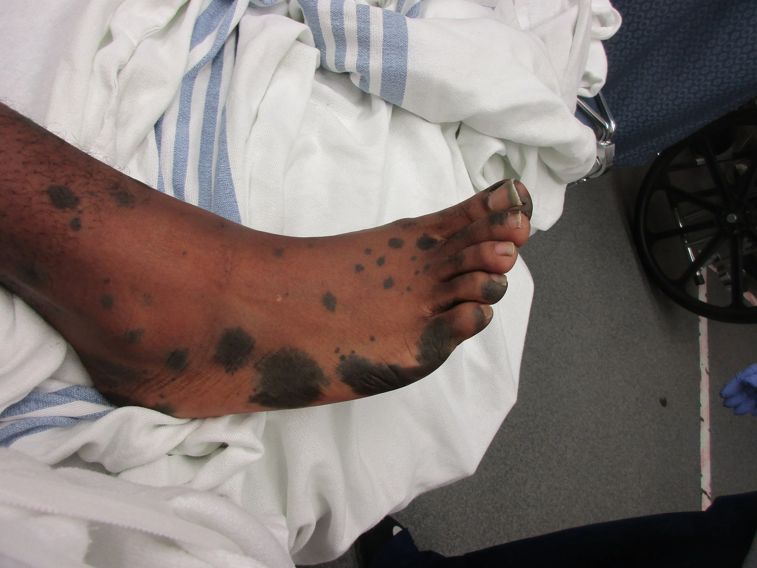
Our patient had neurocutaneous melanosis. This is a rare disease characterized by pigmented nevi that affect not only the skin but the leptomeninges. It was first described in 1861 by Rokitanski. About 1/3 of the patients diagnosed are symptomatic often presenting with seizures, cranial nerve palsies, hydrocephalus or spinal cord involvement. It is associated with Dandy-Walker malformation in 10% of the cases. Our patient had presented with seizures as a child and on imaging was found to have melanosis in the R temporal lobe. Because of intractable seizures he underwent a R temporal lobectomy in 2015.
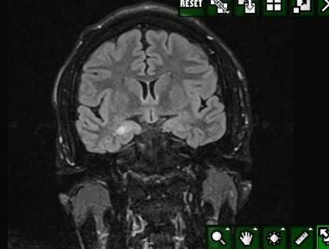
melanosis in the R temporal lobe in our patient prior to surgery
The diagnosis is made on MRI with the melanocytes being hyperintense on T1 images. The deposits of melanocytes are most often seen in the amygdala, cerebellum and upper cervical cord. The pathogenesis is believed to be dysplasia of the neuroectodermal melanocyte precursor cells leading to proliferation of melanin in the skin and leptomeninges. Patients with neurocutaneous melanosis are at risk for malignant transformation to melanoma. Malignant transformation occurs in 40-60% of symptomatic cases.
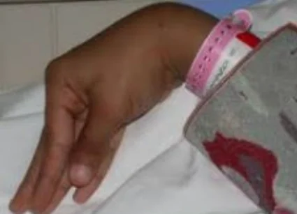
carpopedal spasm
Carpopedal spasm occurs when acute hypocarbia causes reduced ionized calcium and phosphate levels resulting in involuntary contraction of the feet and hands. This can occur with dehydration, hyperventilation (from any cause including MI, infection or bleeding), hypothyroidism, tetanus, and brain disorders (Parkinsons, MS, dystonia and huntingtons).
Our patient had normal Mg, Ca and phosphorus. His CO2 was 20. The cause for his carpopedal spasm was thought to be dehydration from a night of drinking preceding his presentation. He had an elevated ddimer and underwent a PE protocol CT which showed no PE but a small R to L shunt. His spasms resolved with hydration.
Gocmen R, Guler E, Arslan E. A case of neurocutaneous melanosisand neuroimaging findings. 2015 Journal of Radiology Case Reports. 9(3) 1-6.
Rokitanski J, Ein ausgezeichneter Fall von Pigment-Mal mit ausgebreiteter Pigmentierung der inneren Hirn-und Ru chenmarkhaute. Ally Wien Med Z 1861(6):113-16.
Ginat DT, Meyers SP. Intracranial lesions with high signal intensity of T1-weighted MR images: differential diagnosis. Radiographics 2012 32(2) :499-516.